

Files
Download Full Text (1.6 MB)
Publication Date
2025
Keywords
oregon, oaa 2025, ppmc
Disciplines
Medical Education | Neurology | Neurosciences
Abstract
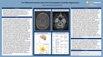
Introduction: Paraneoplastic cerebellar degeneration is a rare cause of cerebellar ataxia with a poor prognosis for neurologic recovery. The management of symptoms is dependent primarily upon timely diag nosis and treatment of the underlying neoplasm. Herein, we present the diagnostic and treatment course of a patient with paraneoplastic cerebellar degeneration. Case: A 72-year-old woman with a past medical history of left mastectomy for high grade ER/PR negative Her2 positive invasive ductal carcinoma presented with progressively worsening dysarthria and ataxia over two months. A metabolic work up by her primary care physician was unrevealing. Breast MRI did not show recurrence of her cancer. During her visit with her oncolo gist, the patient noted a ten-pound weight loss. At her first emergency department visit, exam revealed dysarthria but was an otherwise normal neurologic exam. MRI brain without contrast and MRA head and neck without contrast showed no evidence of ischemia, hemorrhage or mass effect. She was discharged home with the plan to follow up with outpatient neurology. Three days later, the patient presented to another emergency department and was found to be dysarthric but otherwise neurologically intact. CT head without contrast and CTA head and neck did not reveal a structural cause. MRI with and without contrast noted a non-specific T2 FLAIR hyperin tense signal associated with the sulci or surface of the cerebellum. CSF showed elevated protein twice the upper limit of normal and a higher-than-normal fraction of lymphocytes. During her hospitalization she was noted to have worsening dysarthria, bilateral upper and lower extremity ataxia and development of diplopia for which she received 500mg methylprednisolone twice daily for three days and IVIG for five days. Symptoms were initially responsive but progressed further prompting five days of plasmapheresis. Symptoms did not remit. CA-125 was elevated at 253 U/mL and the CSF paraneoplastic panel was positive for anti-Yo antibody at 1:61440. A PET CT detected two para-aortic lymph nodes. Pathology from a total abdominal hysterectomy and bilateral salpingo-oophorectomy, peritoneal washing, and singular lymph node removal yielded carcinoma of mullerian origin in the lymph node alone. Steroids and cyclophosphamide were trialed for persistent neurologic symptoms. She received two cycles of docetaxel and carboplatin. The patient's care was transferred to a facility closer to family. Discussion: Paraneoplastic cerebellar degeneration is a rare cause of subacute cerebellar ataxia with an inci dence of 0.41-0.89/100,000 person years2,3. Associated malignancies include most commonly small cell lung cancer, ovarian cancer, breast cancer and Hodgkin lymphoma. While several auto antibodies have been identified as drivers of cerebellar degeneration, the anti-Yo antibody is predominantly associated with gynecological and breast cancers. Greater than 90% of these can cers identified while alive or on autopsy4. Brain MRI rarely shows cerebellar enhancement, as seen in the described patient, and more typically is normal or shows cerebellar degeneration. Anti-Yo (purkinje cell cytoplasmic antibody type 1) can be detected in serum or CSF. It is the most commonly associated antibody intracellularly targeting the cerebellar degeneration-related 2-like (CDR2L) antigen within the Purkinje cells of the cerebellum6. When bound, it leads to cell death by inhibiting the ribosome to which it is bound. Once the diagnosis of paraneoplastic cerebellar ataxia is suspected, it is important to search for the source of the neoplasm since the most effec tive antidote is treating the primary malignancy. Unfortunately, 75-80% of patients become and remain non-ambulatory with this condition, with delay of diagnosis likely contributing to poor outcomes4. There are anecdotal reports of neurologic improvement with IVIG, plasmapheresis, steroids, azathioprine, cyclophosphamide, and rituximab5. Albeit rare, it is important to be highly suspicious of autoantibody mediated neuronal degenerative diseases as symptom management depends on timely diagnosis and treatment. Conclusion: When work-up for more common causes of cerebellar ataxia is negative and/or if a paraneoplastic source is suspected, imaging and CSF testing can guide further evaluation. Once the diagnosis of para neoplastic cerebellar ataxia is suspected, it is important to search for the source of the neoplasm since the most effective antidote is treating the primary malignancy. Albeit rare, it is important to be highly suspicious of autoantibody mediated neuronal degenerative diseases as symptom management depends on timely diagnosis and treatment
Area of Special Interest
Neurosciences (Brain & Spine)
Specialty/Research Institute
Graduate Medical Education
Specialty/Research Institute
Neurosciences