
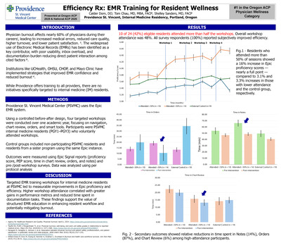
-

Cloudy Consequences: Exploring Vaping-Induced Pulmonary Alveolar Proteinosis
Jennifer Wong, DO; Omar Pandhair; Abby Lindseth, MD; and Cameron Miles Long, MD
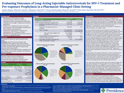
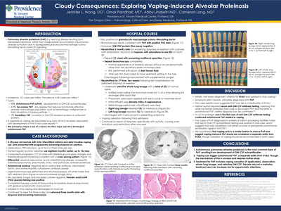
Abstract: Pulmonary alveolar proteinosis (PAP) is a rare disease caused by abnormal accumulation of surfactant within alveoli. Vaping of electronic cigarette smoke has recently been described as a cause of pulmonary alveolar proteinosis in two case reports. We add to this rare literature by describing a case of pulmonary alveolar proteinosis induced from vaping in a young woman with confirmed granulocyte/macrophage colony-stimulating factor (GM-CSF) autoantibodies as the proposed mechanism. Introduction: Pulmonary alveolar proteinosis (PAP) results from inappropriate accumulation of pulmonary alveolar surfactant due to dysregulated granulocyte/ macrophage colony-stimulating factor (GM-CSF) signaling.1 Its incidence is approximately 0.2 cases per million with a prevalence between 4 and 40 cases per million.2 Autoimmune PAP (aPAP) due to development of anti-GM-CSF antibodies accounts for 90% of cases and the remaining 10% arise mainly from secondary causes such as hematologic malignancies or toxic inhalation of mineral particles.1,2 E-cigarette or vaping-use associated lung injury (EVALI) has been associated with development of PAP in two case reports.3,4 We present a unique case of a heavy nicotine vape user who developed autoimmune PAP. Case Presentation: A 28-year-old woman with mild, intermittent asthma presented to emergency department with subacute cough, dyspnea, and mild hypoxemia. Eighteen months earlier, she began regularly using a nicotine vaporizer fifteen times daily. She was mildly tachycardic with diffuse crackles on pulmonary auscultation. A computed tomography (CT) scan of the chest revealed bilateral ground-glass changes and interlobular septal thickening consistent with a crazy paving pattern (Fig. 1A). The initial differential diagnosis included atypical pneumonia, acute interstitial lung disease, pneumonitis, EVALI, and pulmonary alveolar proteinosis. Autoimmune workup including serum antinuclear antibody, rheumatoid factor, and antineutrophilic cytoplasmic antibody panels were negative. Urgent bronchoscopy was performed and was notable for the return of opaque, off-white fluid on bronchoalveolar lavage (BAL). While BAL bacterial, fungal, and Nocardia cultures returned negative, periodic acid-Schiff (PAS) special staining was positive. She finished a five-day course of Doxycycline for possible atypical pneumonia with gradual symptomatic improvement. She was advised to stop vaping and discharged on room air. The patient continued to vape five times a day and returned three months later with dyspnea and worsening hypoxemia. A repeat CT scan showed worsening multifocal opacities and increased septal thickening (Fig. 1B). She underwent repeat BAL and transbronchial biopsy with pathology and cytology confirming the diagnosis of PAP. Serum GM-CSF was low (Mayo Clinic Laboratories) and serum anti-GM-CSF autoantibodies (National Jewish Laboratories) returned elevated, consistent with autoimmune PAP. She was discharged on supplemental oxygen with exertion and underwent whole lung lavage two weeks later. The lungs were sequentially lavaged (right then left) with 0.9% warm saline (37 degrees Celsius) 1 L at time and allowing gravity to drain. Drainage was aided by a manual chest percussion device. The right lung was lavaged with 12 L and the left lung was lavage with 11.5 L. The lavage effluent progressively became more translucent with each lavage for both lungs (Fig. 2). She was monitored in the ICU overnight and discharged the next morning. Discussion: Initially, our patient met every diagnostic criterion for EVALI and was advised to stop vaping.5 Her symptoms did not resolve and she was ultimately diagnosed with PAP. Two case reports have suggested PAP can be a complication of EVALI. Unfortunately, neither author reported serum anti-GM-CSF antibody testing (National Jewish Laboratories), meaning that while the former attributed their case to secondary PAP and the latter to autoimmune PAP, neither diagnosis can be confirmed.3,4 Israel, AK et.al. 2020 suggested that vape inhalation acts like a toxic inhalation causing direct damage to macrophages, while Chua, TH et. al. 2021 posited that “vaping products ... incite self-reactivity,” to trigger aPAP. To our knowledge, ours is the first case of anti-GM-CSF antibody testing confirmed autoimmune PAP related to vaping. Interestingly, two cases of PAP diagnosed in workers at indium processing facilities noted that serum GM-CSF autoantibody testing was positive in one case, which suggests that toxic inhalations can either uncover or precipitate autoimmune sensitization against GM-CSF.6 We hypothesize that vaping acts in a similar fashion to induce PAP and suggest vaping-induced PAP should be considered a separate entity from EVALI, though cessation of vaping should be recommended in both cases. Treatment for PAP can involve observation, whole lung lavage, and nebulized GM-CSF.7 Steroids have not been proven effective and can increase the risk of opportunistic infections. Given the patient’s initial improvement, vaping cessation alone was recommended. A steroid trial was considered as it seemed possible that she could have both EVALI and pulmonary alveolar proteinosis, but this was not initiated. The patient continued to vape and had further deterioration of her lung function which ultimately led to the whole lung lavage. The patient stopped vaping after the whole lung lavage and was started on nebulized GM-CSF.
-

Hepatic Schistosomiasis: An Unexpected Culprit Behind the Hepatic Mass
Jin Yun DO; Udip Dahal MD; and Kenneth Flora, MD
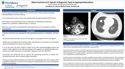
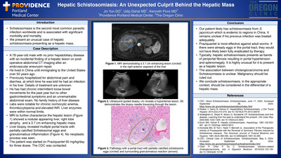
Introduction: Schistosomiasis is the second most common parasitic infection worldwide and is associated with significant morbidity and mortality. We present an unusual case of hepatic schistosomiasis presenting as a hepatic mass. Case Description: A 78-year-old male with no prior hepatobiliary disease with an incidental finding of a hepatic lesion on postoperative abdominal CT imaging after an endovascular aneurysm repair. He lived in China until immigrating to the United States over 50 years ago. Previously hospitalized for abdominal pain and diarrhea, at which time he was told he had an infection in his liver. Details of treatment are unknown. He has had chronic intermittent loose bowel movements for the past year but no other gastrointestinal symptoms and an unremarkable abdominal exam. No family history of liver disease. Labs were notable for chronic normocytic anemia, thrombocytopenia and elevated INR. Liver enzymes were within normal limits. MRI to further characterize the hepatic lesion showed a nodular appearing liver, right lobe atrophy, and a 3.7 cm enhancing hepatic mass. Liver biopsy revealed multiple portal tracts with partially calcified Schistosomal eggs and granulomatous inflammation. No Neoplastic process was seen. The patient was started on Praziquantel 60 mg/kg/ day for three doses. The CDC was contacted. Conclusion: Our patient likely has schistosomiasis from S. japonicum which is endemic to regions in China. It remains unclear if his previous infection was treated adequately. Praziquantel is most effective against adult worms. If there were already eggs in the portal tract, they would not have likely been fully eradicated by therapy. Typically, hepatic schistosomiasis presents as bands of periportal fibrosis resulting in portal hypertension and splenomegaly. It is highly unusual for it to present as a hepatic lesion. The association between cholangiocarcinoma and Schistosomiasis is unclear. Malignancy should be ruled out. We conclude schistosomiasis, in the appropriate context, should be considered in the differential of a hepatic mass.
Printing is not supported at the primary Gallery Thumbnail page. Please first navigate to a specific Image before printing.


