
Files
Download Full Text (827 KB)
Publication Date
4-29-2026
Keywords
oregon, psvmc, psvmc gme, psvmc oaa, psvmc pharmacy gme
Disciplines
Internal Medicine | Medical Education
Abstract
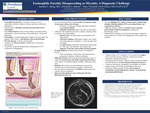
Introduction: Eosinophilic fasciitis (EF), or Shulman’s disease, is a rare disorder of unknown etiology and poorly understood pathogenesis. It is characterized by fibroblast activation and interleukin-driven eosinophilia. Early clinical features include limb or trunk erythema, non-pitting edema, and progressive fascial thickening, often producing an “orange peel” appearance. Eosinophilia is a prominent laboratory finding in the early phase, but diagnosis often requires a full-thickness skin-to-muscle biopsy. Systemic corticosteroids are first-line therapy, with methotrexate as the steroid -sparing agent for refractory disease. We present a unique and diagnostically challenging case of EF confirmed by histopathology. Case Presentation: A 56-year-old man presented with two-months of progressive right forearm pain, edema, and erythema refractory to conservative therapy and empiric antibiotics for presumed cellulitis. An outpatient ultrasound excluded deep vein thrombosis (DVT) and worsening symptoms prompted hospital admission. Examination revealed non-pitting edema and induration of the right forearm; inflammatory markers were normal and mild peripheral eosinophilia (1.5 times the upper limit of normal) was noted. CT showed diffuse subcutaneous edema along the volar aspect of his right forearm. Necrotizing fasciitis and compartment syndrome were considered but deemed unlikely given benign clinical findings by General and Orthopedic Surgery. His workup included evaluation for myositis, eosinophilic fasciitis, and scleroderma, among other diagnoses. MRI demonstrated fascial edema and thickening, resulting in a muscle biopsy and single compartment fasciotomy with evidence of immediate bulging of healthy underlying muscle tissue. Histology showed dense mixed inflammation in perimysial connective tissue with prominent eosinophils, confirming EF. Lastly, his myositis panel returned positive for PM-SCL antibodies. The patient improved on a prednisone taper and was discharged with close Rheumatology follow-up. Discussion: Though the etiology of EF is unknown, it is associated with triggers such as strenuous exercise, infections, autoimmune diseases, medications, and physical stressors. Autoimmune thyroid disease, Sjogren’s, lupus, rheumatoid arthritis, and scleroderma are the most associated autoimmune diseases to EF. Cutaneous involvement, neuropathies, and myalgias are common, though visceral involvement and arthropathies may occur. Laboratory findings typically include early peripheral eosinophilia, elevated inflammatory markers, and polyclonal hypergammaglobuline mia. This case highlights EF’s ability to mimic urgent, localized limb etiologies including cellulitis, DVTs, necrotizing fasciitis, and compartment syndrome. Additional etiologies should be considered when symptoms persist despite standard management, including rare rheumatologic causes. Detailed history, attention to evolving data, and iterative diagnostic reassessment were essential in the management of this complex rare condition.
Specialty/Research Institute
Graduate Medical Education
Specialty/Research Institute
Pharmacy
Specialty/Research Institute
Internal Medicine


