
Files
Download Full Text (1.3 MB)
Publication Date
4-29-2026
Keywords
oregon, ppmc, ppmc gme
Disciplines
Medical Education
Abstract
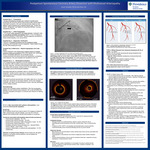
Case Presentation: Hospital Day 0 – Presentation: 43-year-old postpartum female (s/p C-section 4-months prior) presenting with recurrent central chest pressure after several days of intermittent symptoms. She was hemodynamically stable but had diffuse T‑wave inversions and a marked troponin rise. Treated with aspirin, nitroglycerin, and heparin for presumed NSTEMI. Coronary angiography planned. Hospital Day 1 -- Initial Angiography: TTE showed mildly reduced EF with apical akinesis. Angiogram read as mild diffuse mid-distal LAD disease (Figure 1), but no evidence of obstructive disease or culprit lesion. Working diagnosis was MINOCA, stress cardiomyopathy, or an occult SCAD. Hospital Day 2 -- Diagnostic Clarification: Because of persistent symptoms and her postpartum status, cardiology recommended a second angiogram. Patient stable through the day while awaiting the study. Hospital Day 2 (Afternoon) -- Repeat Angiography and Code Event: Repeat angiogram with intracoronary imaging (Figure 2) revealed extensive intramural hematoma of the mid‑to‑apical LAD, confirming SCAD. Immediately afterward, she developed abrupt unresponsiveness with seizure activity and hypotension, prompting a code response. She was intubated and transferred to the ICU. Hospital Day 2 - 4 -- Multisystem Evaluation CVA imaging showed irregularity of the left vertebral artery concerning for dissection. MRI revealed a small acute cerebellar infarct. CTA of the chest, abdomen, and pelvis demonstrated small‑caliber vessels and celiac ostial narrowing, raising concern for an underlying arteriopathy such as fibromuscular dysplasia. Neurology attributed the cerebellar infarct to vertebral artery dissection. Cardiac function improved on repeat echocardiography, supporting stress cardiomyopathy (likely precipitated by SCAD) rather than ischemic injury. The patient remained asymptomatic with stable vitals and no recurrent chest pain. Remained at neurologic baseline. SCAD was managed medically with aspirin and blood‑pressure control. She was discharged home in stable condition. Post-discharge: Patient has completed cardiac rehabilitation. She has no long-term deficits from left vertebral artery dissection. BP is well-controlled with low-dose amlodipine and aspirin is continued with intention for lifelong treatment. Takeaways: SCAD is a distinct cause of MI It is non-atherosclerotic and non-traumatic, often affecting patients without traditional cardiovascular risk factors, requiring a management approach different from typical ACS. SCAD often reflects systemic arteriopathy A high percent of patients have extra coronary vascular abnormalities, most commonly fibromuscular dysplasia, supporting head-to-pelvis vascular imaging at time of diagnosis Angiography can be misleading, and intracoronary imaging is key in these cases Type 2 and Type 3 SCAD can mimic atherosclerosis. OCT clarifies the diagnosis by demonstrating true lumen compression and intramural hematoma, as in this case. SCAD management prioritizes vessel healing, symptom control, and recurrence prevention rather than routine revascularization Most SCAD lesions heal spontaneously with conservative therapy. Conservative management is preferred in stable patients PCI carries higher risk of dissection extension and lower success rates, and is reserved for ongoing ischemia, hemodynamic instability, or high‑risk anatomy. Medical therapy targets SCAD pathophysiology Aspirin is commonly used to reduce superimposed thrombotic risk after MI. Anticoagulation is generally discontinued once SCAD is confirmed. Beta blockers reduce arterial shear stress and are associated with lower recurrence Post‑SCAD chest pain is common. Persistent symptoms may reflect vasospasm or microvascular dysfunction, and CCBs and nitrates are useful for symptom control. Recurrence risk is high. SCAD re-occurs in approximately 5-15% of patients over 2-5 years, typically in a different coronary vessel, reinforcing the focus on prevention rather than revascularization.
Specialty/Research Institute
Graduate Medical Education
Specialty/Research Institute
Internal Medicine


