
-
A Vexing Case: A Newly Discovered Autoinflammatory Disease
Ashley Edwards, MD and Caroline McCulley
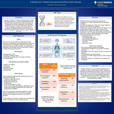
Introduction: Vacuoles, E1 enzyme, X-linked, autoinflammatory, somatic (VEXAS) Syndrome is a recently recognized adult-onset autoinflammatory disease that can lead to potentially lifethreatening multi-system inflammation with estimated prevalence of up to 1 in 5,000 males over the age of 50. Given its significant morbidity and mortality and high prevalence, increased awareness of this condition is of foremost importance. Herein we outline a case of VEXAS syndrome. Case Presentation: Mr. N is a 73-year-old male with a past medical history significant for hypertension and recent episode of uveitis who first presented to the hospital for significant unintentional weight loss, fevers, and abdominal pain. Two weeks later he presented to the ED again due to recurrent fevers, nausea, vomiting, sore throat, and eye pain. Given this multi-system inflammatory disease of unclear etiology, VEXAS syndrome was considered. The UBA1 genetic test was obtained and positive, confirming the diagnosis. He was started on high dose of steroids with a slow taper and remains in remission on 5mg of prednisone daily. Discussion: VEXAS syndrome has a broad phenotype and can masquerade as multiple inflammatory and hematological conditions. The differential diagnosis is nj8broad and includes malignancy, infections, and autoimmune conditions. Prevalence is as high as 1/4269 men over the age of 50, with mortality rates as high as 50% within 4 years of onset. Multiple reviews suggest that VEXAS ought to be heavily considered in men >50 years old presenting with multisystem inflammatory disease with hematological abnormalities. Common presenting symptoms include: • Noninfectious fever • Unintentional weight loss • Dermatological manifestations • Pulmonary infiltrates • Chondritis • Macrocytic anemia Diagnosis and Treatment: Bone marrow biopsy typically demonstrating myelodysplastic changes and vacuolization of myeloid precursors. Identification of the UBA1 genetic mutation is necessary for diagnosis. Initial treatment is high dose steroids. Consider IL-6 inhibitor biologic therapy if unable to taper off Prednisone. Patients are typically co-managed by rheumatology and hematology
-
The Pernicious Right Should Pain
Julie Hwang, MD; Weiya Wysham, MD; and Qian Leng, MD
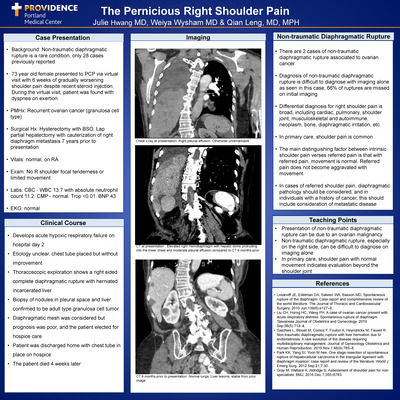
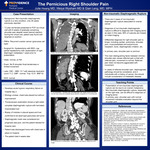
Case Presentation: •Background: Non-traumatic diaphragmatic rupture is a rare condition, only 28 cases previously reported •73 year old female presented to PCP via virtual visit with 6 weeks of gradually worsening shoulder pain despite recent steroid injection. During the virtual visit, patient was found with dyspnea on exertion •PMHx: Recurrent ovarian cancer (granulosa cell type) •Surgical Hx: Hysterectomy with BSO, Lap partial hepatectomy with cauterization of right diaphragm metastasis 7 years prior to presentation •Vitals: normal, on RA •Exam: No R shoulder focal tenderness or limited movement •Labs: CBC - WBC 13.7 with absolute neutrophil count 11.2. CMP - normal. Trop < 0.01. BNP 43 •EKG: normal Discussion: •There are 2 cases of non-traumatic diaphragmatic rupture associated to ovarian cancer •Diagnosis of non-traumatic diaphragmatic rupture is difficult to diagnose with imaging alone as seen in this case, 66% of ruptures are missed on initial imaging •Differential diagnosis for right shoulder pain is broad, including cardiac, pulmonary, shoulder joint, musculoskeletal and autoimmune, neoplasm, bone, diaphragmatic irritation, etc. •In primary care, shoulder pain is common •The main distinguishing factor between intrinsic shoulder pain verses referred pain is that with referred pain, movement is normal. Referred pain does not become aggravated with movement •In cases of referred shoulder pain, diaphragmatic pathology should be considered, and in individuals with a history of cancer, this should include consideration of metastatic disease Teaching Points: •Presentation of non-traumatic diaphragmatic rupture can be due to an ovarian malignancy •Non-traumatic diaphragmatic rupture, especially on the right side, can be difficult to diagnose on imaging alone •In primary care, shoulder pain with normal movement indicates evaluation beyond the shoulder joint
-
Case Study: A Severe Presentation of Chronic Eosinophilic Pneumonia.
Revati Kalluri and Mari Kai
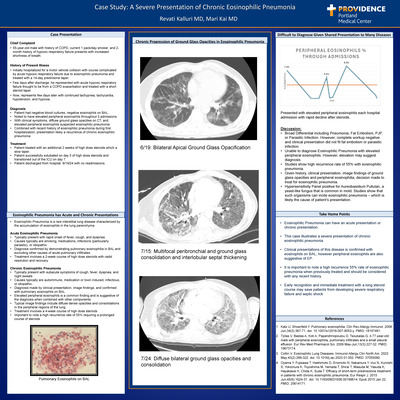
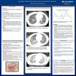
Case Presentation: Chief Complaint •55-year-old male with history of COPD, current 1 pack/day smoker, and 2-month history of hypoxic respiratory failure presents with increased shortness of breath. History of Present Illness •Initially hospitalized for a motor vehicle collision with course complicated by acute hypoxic respiratory failure due to eosinophilic pneumonia and treated with a 14-day prednisone taper. •Few days after discharge, he represented with acute hypoxic respiratory failure thought to be from a COPD exacerbation and treated with a short steroid taper. •Now, represents few days later with continued tachypnea, tachycardia, hypotension, and hypoxia.. Discussion: •Broad Differential including Pneumonia, Fat Embolism, PJP, or Parasitic Infection. However, complete workup negative and clinical presentation did not fit fat embolism or parasitic infection. •Unable to diagnose Eosinophilic Pneumonia with elevated peripheral eosinophils. However, elevation may suggest diagnosis. •Studies show high recurrence rate of 55% with eosinophilic pneumonia. •Given history, clinical presentation, image findings of ground glass opacities and peripheral eosinophilia, decision made to treat for eosinophilic pneumonia. Hypersensitivity Panel positive for Aureobasidium Pullulan, a yeast-like fungus that is common in mold. Studies show that such organisms can incite eosinophilic pneumonia – which is likely the cause of patient’s presentation Take Home Points; •Eosinophilic Pneumonia can have an acute presentation or chronic presentation. •This case illustrates a severe presentation of chronic eosinophilic pneumonia. •Clinical presentations of this disease is confirmed with eosinophils on BAL, however peripheral eosinophils are also suggestive of EP. •It is important to note a high recurrence 55% rate of eosinophilic pneumonia when previously treated and should be considered with any recent history. •Early recognition and immediate treatment with a long steroid course may save patients from developing severe respiratory failure and septic shock
-
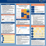
From Dormant to Dangerous: Hepatitis B Reactivation Under Immunosuppression
Adam Knott, MD and Amy Dechet
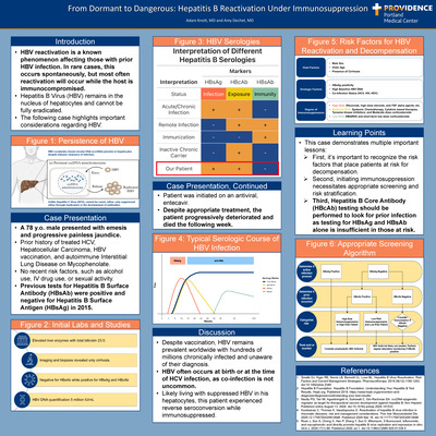
Introduction: •HBV reactivation is a known phenomenon affecting those with prior HBV infection. In rare cases, this occurs spontaneously, but most often reactivation will occur while the host is immunocompromised. •Hepatitis B Virus (HBV) remains in the nucleus of hepatocytes and cannot be fully eradicated. •The following case highlights important considerations regarding HBV. Case Presentation: •A 78 y.o. male presented with emesis and progressive painless jaundice. •Prior history of treated HCV, Hepatocellular Carcinoma, HBV vaccination, and autoimmune Interstitial Lung Disease on Mycophenolate. •No recent risk factors, such as alcohol use, IV drug use, or sexual activity. •Previous tests for Hepatitis B Surface Antibody (HBsAb) were positive and negative for Hepatitis B Surface Antigen (HBsAg) in 2015. Learning Points: •This case demonstrates multiple important lessons: at risk.First, it’s important to recognize the risk factors that place pa tients at risk for decompensation. Second, initiating immunosuppression necessitates appropriate screen ing and risk stratification. Third, Hepatitis B Core Antibody (HBcAb) testing should be performed to look for prior infection as testing for HBsAg and HBsAb alone is insufficient in those
-
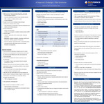
A Diagnostic Challenge—TINU Syndrome
Sophia Lee, MD and Claire Kassakian, MD
Introduction: •TINU Syndrome: Tubulointerstitial Nephritis and Uveitis rare multi-system syndrome has renal manifestations typical of acute interstitial nephritis (AIN), in addition to bilateral anterior uveitis not well-understood •Thought to be an immune-mediated process, although mechanisms for disease remain unclear Likely secondary to a combination of genetic predisposition and certain environmental exposures •Genetic predisposition: specific HLA phenotypes possibly an auto-antigen to modified C-reactive protein (mCRP) •Potential triggers: Infections: tuberculosis, EBV, varicella zoster, chlamydia, toxoplasma Medications: antibiotics (β-lactams, meropenem, azithromycin, levofloxacin), NSAIDs, goreisan (Chinese herb) Endocrine diseases: hypoparathyroidism, hyperparathyroidism, rheumatoid arthritis, IgG4-related autoimmune disease, thrombotic microangiopathy Discussion & Learning Posts: •This case is an example of an extremely rare condition with potentially devastating outcomes if not caught early enough visual impairment dialysis dependence •Treatment for patients with progressive renal dysfunction is usually high-dose prednisone of 1 mg/kg per day (up to 40-60 mg/day) 3-6 month duration of treatment (depending on response) Long taper •Rarely, some patients are treated with mycophenolate mofetil if no benefit from steroid therapy •Fortunately, kidney disease can be self-limited in many patients, however, patients who have developed TINU syndrome are at risk of relapses •Demonstrates the need for a high clinical suspicion for this combination of symptoms: a pyuria suspicious for AIN tubular proteinuria eye redness and pain •Symptoms often do not correlate temporally! In fact, the majority of cases present with renal injury preceding uveitis •important to include TINU in the differential thoroughly assess a review of all systems when taking a history •Also key to note is the risk of future recurrence in patients who have developed TINU syndrome Need for ongoing monitoring
-
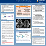
A Deadly Case of Encephalopathy
Brenna Ostertag, MD; Gita D Gelfer; and Andrea Roast
Introduction: Human prion disease are heterogenous groups of neurodegenerative disorders characterized by spongiform degeneration, astrogliosis, and neuronal loss in the central nervous system from misfolding and subsequent spread of the prion protein. This disease manifests rapidly and is often accompanied by visual and speech abnormalities, gait changes, and progressive encephalopathy. It is always fatal. Around 400 people are diagnosed with prion disease in the United States annually. Recent innovations in diagnostics using Real-Time Quaking Induced Conversion (RT-QuIC) has allowed for antemortem diagnosis of prion disease. Case Presentation: A 66-year-old male with a history of alcohol use disorder in remission and tobacco use disorder presented to the emergency department with a broken right humerus after a fall at home. He underwent a successful open reduction internal fixation (ORIF) of his humerus. His hospital course was complicated by encephalopathy and hallucinations, which improved at time of discharge. He presented one week later from his skilled nursing facility with recurrent falls and progressive encephalopathy. At the time of admission, he was oriented to self only. He had multiple complications during this admission including osteomyelitis, vancomycin indued IgA bullous vasculitis, and acute Covid-19 infection. His mentation did not improve throughout his stay and progressively declined despite treatment of comorbidities. Laboratory work-up for causes of his progressive encephalopathy were wholly unremarkable. A lumbar puncture was performed under anesthesia after all other possible causes for his encephalopathy were ruled out. His lumbar puncture demonstrated a positive diagnosis of prion disease. His symptoms progressed to akinetic mutism by the end of his hospital stay. He was discharged to home on hospice and died approximately 4 months after his initial presentation to our hospital. Discussion: Sporadic Creutzfeldt-Jakob Disease (sCJD) accounts for 85% of all human prion disease diagnosed worldwide. The mechanism is believed to be spontaneous conversion of the normal prion proteins to the pathologic variant. This may also be related to an underlying mutation that occurs in the prion gene. Patients typically present initially with falls before symptoms progress to encephalopathy and eventually akinetic mutism prior to death. The patient in this case had a very typical presentation and trajectory for sCJD (we are still awaiting final pathology results from autopsy). The Genetics of Human Prion Diseases, The prion gene sits on human chromosome 20. This is the basis of presumed genetic mutations for genetic causes of human prion disease and possible sCJD. In experimental animal models, the pathologic variants replicate in peripheral lymphoid tissue before entering the CNS. Final transportation of the pathologic prion protein travels to the central nervous system in a retrograde fashion via axons. Techniques for Antemortem Diagnosis RT-QuIC has been studied for over 10 years regarding viability for diagnosis of prion diseases and was added to the diagnostic criteria in 2018. Turn around can be quick, with results becoming available in 3-4 days. The process, as demonstrated in Figure 2, involves the seeding nucleation hypothesis. CSF is obtained by lumbar puncture and is placed in assays and tagged with thioflavin so converted proteins can be seen under fluorescence. MRI findings and EEG findings can be supportive but are not specific. 14- 3-3 protein in the CSF is also considered non-specific but is generally elevated in human prion diseases. Future Directions in Diagnosis and Treatment. Other matrixes besides CSF are being considered. Olfactory mucosa has shown similar sensitivity and specificity to CSF. Other matrixes that have been tested include muscle and skin. Treatment will remain a challenge and will likely require genetic therapy, such as CRISPR, however immunotherapies are also being explored.
-

Assessing Disparities in Evaluation of Heavy Menstrual Bleeding in Patients with Iron Deficiency
Nidhi Patel, MD and Bethany Samuelson Bannow
Background: • Iron deficiency (ID) rates are high in lower income populations and minoritized groups • IDA is often underrecognized and undertreated • Heavy menstrual bleeding is a common yet unrecognized cause for iron deficiency anemia amongst menstruating patients Discussion: • White patients with iron deficiency have less documentation of menstruation than non-white patients • Only about half of all patients have documentation of menstruation or receive additional workup • Of the charts that mention menstruation, most do not document additional details for duration, frequency between periods, and number of products used • Using a questionnaire set (figure 1) may provide more clarity on light versus "normal" versus heavy periods • Referrals to gynecology or hematology are often underutilized Conclusion: • More thorough discussions of menstruation may provide more diagnostic clarity for iron deficiency • ID is always secondary to an underlying cause, so further workup may be warranted
-
When Calciphylaxis isn’t the Answer: Diagnostic Delay in an End Stage Renal Disease Patient with Severe Necrotic Wounds
Megan Schermerhorn, MD; Caroline McCulley; and Courtland Childers
-
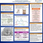
A potentially Cat-astrophic Missed Diagnosis: Systemic Autoimmune Disease or Something more Sinister
Cameron Smith, DO, MPH and Amy Dechet
Introduction: Disseminated bartonella infection has a mortality rate approaching 10%. Clinical presentation of bartonellosis can mimic systemic autoimmune disease. Traditional testing may not identify the organism, so a high degree of suspicion is warranted in patients with risk factors. Case Presentation: 32M with history of Tetralogy of Fallot with prosthetic pulmonary valve presented with fatigue x3 months. Reported severe splenomegaly, pancytopenia requiring transfusions, and renal failure requiring hemodialysis. Given concern for lupus-like syndrome, treated with steroids + cyclophosphamide. Persistent fevers despite broad antibiotics. Second opinion of renal biopsy: Postinfectious GN. Immunostains ruled out lupus-like disorder. Negative blood cultures, TEE, and serologies for HIV, EBV, CMV, hepatitis, coxiella, brucella, and bartonella. Takeaways: • Disseminated bartonellosis can mimic autoimmune diseases. • Patients with exposure to cats and with valvular heart disease are at highest risk. • False negatives common with serologies, blood cultures, and traditional PCR. • In patients with higher indices of suspicion, enhanced PCR’s at specialty labs, valve cultures, ddPCR, or cell-free DNA testing can enhance sensitivities.
-
Endocarditis Prophylaxis in Vulnerable Valve; Antibiotic Stewardship or Suboptimal Care?
Zachary Taylor, Andrea Roast, and Michael Layoun
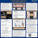
Introduction Infective endocarditis (IE) is a relatively uncommon disease with an annual incidence of 5 per 100,000 people in the U.S.. Dental procedures are an exceptionally rare cause of IE. Amidst a global emphasis on antibiotic stewardship, U.S. and European guidelines only recommend antibiotic prophylaxis for dental procedures in high-risk groups. Having a bicuspid aortic valve (BAV) confers an 11-fold risk of endocarditis compared to the general public, and yet, is not classified as high-risk per AHA/IDSA guidelines. We present a patient with BAV who developed IE following a noninvasive dental procedure and review whether patients with BAV are under-classified and warrant antibiotic prophylaxis. Case presentation A 58-year-old previously healthy male presented to the ED with a 4-6-week history of fatigue, progressive dyspnea, and intermittent fevers. Two weeks prior to onset of symptoms, he underwent a routine dental cleaning. Examination revealed decreased bibasilar breath sounds, pitting edema to the mid-shins, a new diastolic murmur, and bounding carotid pulses. Work-up revealed leukocytosis, elevated procalcitonin, and small bilateral pleural effusions. Cefepime was started for presumed sepsis. Admission blood cultures grew Aggregatibacter aphrophilus, a HACEK organism. Transthoracic echocardiogram demonstrated a focal vegetation of the aortic valve, confirming bacterial endocarditis. There was moderate-to-severe aortic insufficiency (AI), though concomitant severe left ventricular enlargement suggested a degree of chronic AI. Urgent bioprosthetic aortic valve replacement was performed on hospital day 7. Pathology of the excised aortic valve demonstrated bicuspid morphology with Sievers type 2 anatomy. The patient recovered well after 6 weeks of ceftriaxone therapy. Discussion The patient’s HACEK bacteremia and endocarditis were attributed to his recent noninvasive dental cleaning. The diagnosis raised concern for underlying valvular pathology, and the patient was found to have BAV, which is the most common congenital cardiac abnormality and present in approximately 2% of the population. Native valve endocarditis following dental procedures is rare, with an estimated incidence of 1 case per 14 million procedures. The American Heart Association Prevention of Endocarditis guidelines only recommend antibiotic prophylaxis for odontologic procedures in high-risk cardiac conditions. This currently excludes BAV, though newer retrospective studies suggest that patients with BAV may be at higher risk of IE than previously thought. For example, a study by Zegri-Reiriz et al. showed that patients with BAV had a higher incidence of viridans group streptococcus IE and IE from suspected odontologic origin compared to traditionally high-risk groups (14.8% vs. 5.8%; p< 0.01). Additionally, BAV-related IE often leads to perivalvular abscesses, requiring earlier surgery in nearly 75% of cases. Both this case and recent studies question whether BAV warrants reclassification as a high-risk condition for IE. Additional studies are required to further explore the benefit of antibiotic prophylaxis in this group.
-
Evaluation of extended-interval thyroid function monitoring during pembrolizumab treatment
Hanna Yoon, PharmD and Heather Beugli
-
From Hormones to Pancreatitis: A Case of Estrogen-Induced Hypertriglyceridermia
Kyaw Zin (Kennis) Htet, MD; Michael Lefor, MD; and B C. Clark, MD
Introduction: Hormonal replacement therapy (HRT) for transgender women, also known as gender-affirming hormone therapy (GAHT), is designed to align physical characteristics with gender identity. Potential risk associated with feminizing hormone therapy with estradiol include venous thromboembolism, cardiovascular disease and changes in lipid profile. We present a case of estrogen replacement therapy induced hypertriglyceridemia causing acute pancreatitis in a young transgender woman. Case Presentation: A 30-year-old transgender woman on estrogen and progesterone replacement therapy, as well as history of hypertension and MELAS syndrome admitted with 2-day history epigastric pain, nausea, and decreased urination. Notably, her estradiol dosage had recently increased from 6 mg to 8 mg five months prior. Discussion: Acute pancreatitis is a potentially life-threatening condition with significant morbidity and mortality, often resulting from gallstones or alcohol use. Hypertriglyceridemia, though a less common cause, can trigger pancreatitis by increasing free fatty acid production, leading to pancreatic inflammation and injury. The underlying mechanisms of hypertriglyceridemia-induced pancreatitis are not fully understood but are believed to involve hydrolysis of triglycerides into toxic free fatty acids, causing direct pancreatic injury, endothelial damage, calcium overload, and a pronounced inflammatory response. Estrogen therapy, particularly oral formulations, has been associated with elevated plasma triglyceride levels due to its effects on hepatic lipid metabolism. In this case, estrogen-induced hypertriglyceridemia was the likely precipitant of acute pancreatitis, exacerbated by the patient’s pre-existing conditions, including MELAS syndrome and hypertension. The increase in estradiol dosage may have further contributed to the rise in triglyceride levels. Clinicians should routinely monitor triglyceride levels in transgender women receiving estrogen therapy, especially in those with predisposing conditions. Transdermal estradiol, as used in this patient post-discharge, may pose a lower risk of hypertriglyceridemia due to reduced first-pass hepatic metabolism. Early intervention, lipid control, and switching to safer formulations can mitigate the risk of serious complications like pancreatitis.
-
A Case of Kaposi's Sarcoma with Dyspnea
Claire Higgins, MD and Brinton Clark, MD, MPH
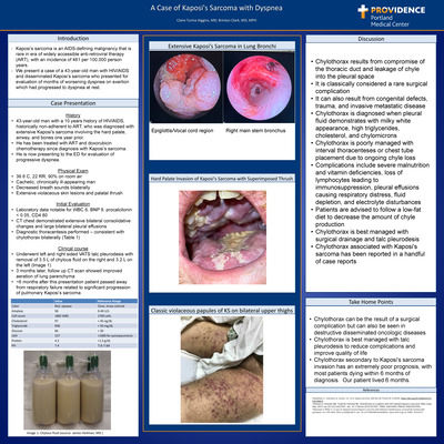
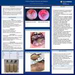
Introduction: Kaposi’s sarcoma is an AIDS-defining malignancy that is rare in era of widely accessible anti-retroviral therapy (ART), with an incidence of 481 per 100,000 person years. We present a case of a 43-year-old man with HIV/AIDS and disseminated Kaposi’s sarcoma who presented for evaluation of months of worsening dyspnea on exertion which had progressed to dyspnea at rest. Case Presentation: •43-year-old man with a 10 years history of HIV/AIDS, historically non-adherent to ART, who was diagnosed with extensive Kaposi's sarcoma involving the hard palate, airway, and bones one year prior. •He has been treated with ART and doxorubicin chemotherapy since diagnosis with Kaposi’s sarcoma. •He is now presenting to the ED for evaluation of progressive dyspnea. Discussion: •Chylothorax results from compromise of the thoracic duct and leakage of chyle into the pleural space •It is classically considered a rare surgical complication •It can also result from congenital defects, trauma, and invasive metastatic disease •Chylothorax is diagnosed when pleural fluid demonstrates with milky white appearance, high triglycerides, cholesterol, and chylomicrons •Chylothorax is poorly managed with interval thoracenteses or chest tube placement due to ongoing chyle loss •Complications include severe malnutrition and vitamin deficiencies, loss of lymphocytes leading to immunosuppression, pleural effusions causing respiratory distress, fluid depletion, and electrolyte disturbances •Patients are advised to follow a low-fat diet to decrease the amount of chyle production •Chylothorax is best managed with surgical drainage and talc pleurodesis •Chylothorax associated with Kaposi's sarcoma has been reported in a handful of case reports
Printing is not supported at the primary Gallery Thumbnail page. Please first navigate to a specific Image before printing.