
-

Legionella: an infectious cause of rhabdomyolysis-associated acute renal failure
Alexander Hall, MD and Meera Jain
-
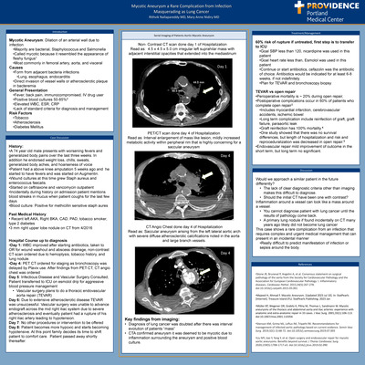
Mycotic Aneurysm and Rare Complication from Infection Masquerading as Lung Cancer
Rithvik Nallapareddy and Mary Anne Nidiry
Introduction: Mycotic Aneurysm: Dilation of an arterial wall due to infection • Majority are bacterial, Staphylococcus and Salmonella • Called mycotic because it resembled the appearance of fleshy fungus1 • Most commonly in femoral artery, aorta, and visceral Causes • Form from adjacent bacteria infections • Lung, esophagus, endocarditis • Direct invasion of vessel walls or atherosclerotic plaque in bacteremia General Presentation • Fever, back pain, immunocompromised, IV drug user • Positive blood cultures 50-85%2 • Elevated WBC, ESR, CRP • Lack of standard criteria for diagnosis and management Risk Factors • Tobacco • Atherosclerosis • Diabetes Mellitus Case Presentation: • A 74 year old male presents with worsening fevers and generalized body pains over the last three weeks. In addition he endorsed weight loss, chills, sweats, generalized body aches, and hoarseness of voice • Patient had a above knee amputation 5 weeks ago and he started to have fevers and was started on Augmentin. • Wound cultures at this time grew Staph aureus and enterococcus faecalis. • Started on ceftriaxone and vancomycin outpatient • Incidentally during history on admission patient mentions blood streaks in mucus when patient coughs for the last few days • Blood culture: Positive for methicillin sensitive staph aures Discussion: Would we approach a similar patient in the future differently? • The lack of clear diagnostic criteria other than imaging makes this difficult to diagnose. • Should the initial CT have been one with contrast? Inflammation around a vessel can look like a mass around a vessel. • You cannot diagnose patient with lung cancer until the results of pathology come back. • A primary lung nodule if found incidentally on CT many years ago likely did not become lung cancer This case shows a rare complication from an infection that requires complex and urgent medical management that can present in an incidental manner • Really difficult to predict manifestation of infection or sepsis around the body.
-
Walking the Razor’s Edge: A Crossover Between Infection and Malignancy
Brenna Ostertag, MD and Gita D Gelfer
-
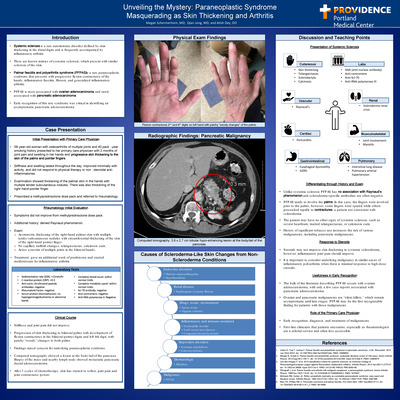
Unveiling the Mystery: Paraneoplastic Syndrome Masquerading as Skin Thickening
Megan Schermerhorn, MD; Quian Leng; and Atish Dey
Introduction: Systemic sclerosis is a rare autoimmune disorder defined by skin thickening in the distal digits and is frequently accompanied by inflammatory arthritis. There are known mimics of systemic sclerosis, which present with similar sclerosis of the skin. Palmar fasciitis and polyarthritis syndrome (PFPAS) is a rare paraneoplastic syndrome that presents with progressive flexion contractures of the hands, inflammatory fasciitis, fibrosis, and generalized inflammatory arthritis. PFPAS is most associated with ovarian adenocarcinoma, and rarely associated with pancreatic adenocarcinoma. Early recognition of this rare syndrome was critical in identifying an asymptomatic pancreatic adenocarcinoma. Case Description: 58-year-old woman with osteoarthritis of multiple joints and 40 pack-year smoking history presented to her primary care physician with 2 months of joint pain and swelling in her hands and progressive skin thickening to the skin of the palms and pointer fingers. Stiffness and swelling lasted throughout the day, improved minimally with activity, and did not respond to physical therapy or non-steroidal anti-inflammatories. Examination showed thickening of the palmar skin in the hands with multiple tender subcutaneous nodules. There was also thickening of the right-hand pointer finger. Prescribed a methylprednisolone dose pack and referred to rheumatology. Discussion: Differentiating through History and Exam - Unlike systemic sclerosis PFPAS has no association with Raynaud’s phenomenon and scleroderma-specific antibodies are often negative. PFPAS tends to involve the palms. In this case, the fingers were involved prior to the palms, however, some fingers were spared while others proceeded rapidly to contractures, a pattern not consistent with scleroderma. The patient may have no other signs of systemic sclerosis, such as severe heartburn, matted telangiectasias, or calcinosis cutis. History of significant tobacco use increases the risk of various malignancies, including pancreatic malignancies. Response to Steroids Steroids may not improve skin thickening in systemic scleroderma; however, inflammatory joint pain should improve. It is important to consider underlying malignancy in similar cases of inflammatory polyarthritis when there is minimal response to high-dose steroids. Usefulness in Early Recognition - The bulk of the literature describing PFPAS occurs with ovarian adenocarcinoma, with only a few case reports associated with pancreatic adenocarcinoma. Ovarian and pancreatic malignancies are “silent killers,” which remain asymptomatic until late stages. PFPAS may be the first recognizable finding for patients with these malignancies.
-
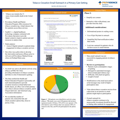
Tobacco Cessation Email Outreach in a Primary Care Setting
Katie Berry, MD and Meera Jain
Background: . • Tobacco use remains the #1 cause of preventable death in the United States • Providence Health and Patient Education Program offers resources for tobacco cessation (eg. Virtual classes), but few patients access these services • Xealth® is a digital healthcare technology company contracted with Providence that aims to provide digital content to improve patient care and resources to analyze data • Study purpose: Assess if digital outreach to patients helps engagement in tobacco cessation services • Our clinic was a pilot clinic to develop the intervention and assess efficacy prior to roll out to all Providence Medical Group clinics in Portland Metro area Methods: • .An email was sent to all patients actively using tobacco at PMG NE Clinic providing information on tobacco cessation and links to click if interested in quitting • The links notified the Health and Patient Education Program to personally call each interested patient to provide cessation resources (virtual behavioral health classes, an office visit in their primary care clinic to discuss tobacco cessation, and/or Oregon Quit Line and American Lung Association information) • We recorded the number of patients who clicked the link and which cessa tion options they chose to pursue if any. Conclusions: • Mass email outreach was not successful in engaging patients in tobacco ces sation • Barriers to participation: • Patients in pre-contemplative stages of quitting • Disregard to automated messages ([email protected]) • Difficulty with email navigation, • Length of email • Modifications to the email style and/or serial messages may result in higher participation rates • Video about tobacco cessation services from a physician in our practice to email patients might improve participation • If email outreach for tobacco cessation continues to be unsuccessful, investment should be directed to finding other methods for engagement
-

Acute Pain Management of Hospitalized Patients on Medication-Assisted Therapies (MAT) for Opioid Use Disorder (OUD)
Ryan Bower, Roland Mikkaeil, Emily H. Hsu, and Tracee Mock
Abstract: Buprenorphine has been an instrumental tool in Medication Assisted Therapies (MAT) for opioid use disorder (OUD). For hospitalized patients on MAT, the management of acute pain presents a unique challenge to health care providers. A previous study examining buprenorphine discontinuation patterns on medical/surgical floors found that 11.8% of patients had their buprenorphine discontinued due to inadequate analgesia. Furthermore, none of these patients had their buprenorphine restarted prior to discharge. Discontinuation of buprenorphine in hospitalized patients has the potential for wide-reaching implications ranging from risk of withdrawal to relapse secondary to patient loss during the transition from inpatient to outpatient care. To improve providers' understanding around buprenorphine and address potential misconceptions regarding its use, educational materials in the form of a presentation aimed towards healthcare providers were developed with guideline-directed strategies to manage hospitalized patients on buprenorphine for OUD. To assess the efficacy of the presentation, pre- and post-surveys were conducted to establish baseline knowledge and determine potential impact on clinical practice. Survey questions were designed using a Likert scale. Responses will be analyzed using a paired T-test. Results and conclusions are currently pending and will be shared upon project completion. (IRB exempt) Learning objectives: Examine the utility and success of education to healthcare providers on analgesic strategies for hospitalized patients on buprenorphine for OUD management. Presentation category: Education/academia/staff development; transitions of care/medication reconciliation; medication safety/quality improvement
-
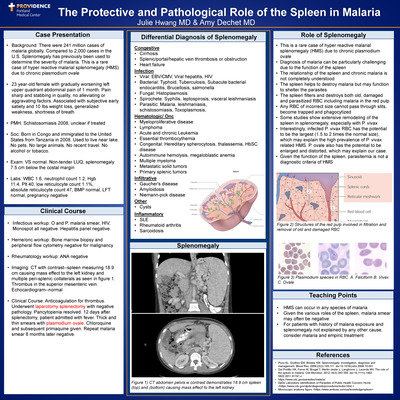
The Protective and Pathological Role of the Spleen in Malaria
Julie Hwang, MD and Amy Dechet
Case Presentation: • Background: There were 241 million cases of malaria globally. Compared to 2,000 cases in the U.S. Splenomegaly has previously been used to determine the severity of malaria. This is a rare case of hyper reactive malarial splenomegaly (HMS) due to chronic plasmodium ovale • 23 year-old female with gradually worsening left upper quadrant abdominal pain of 1 month. Pain sharp and stabbing in quality, no alleviating or aggravating factors. Associated with subjective early satiety and 10 Ibs weight loss, generalized weakness, shortness of breath • PMH: Schistosomiasis 2008,unclear if treated • Soc: Born in Congo and immigrated to the United States from Tanzania in 2008. Used to live near lake. No pets. No large animals. No recent travel. No alcohol or tobacco. • Exam: VS normal. Non-tender LUQ, splenomegaly 7.5 cm below the costal margin • Labs: WBC 1.6, neutrophil count 1.2, Hgb 11.4,Plt40, low reticulocyte count 1.1%, absolute reticulocyte count 47, BMP normal, LFT normal, pregnancy negative Clinical Course: • Infectious workup: O and P, malaria smear, HIV, Monospot all negative. Hepatitis panel negative. • Heme/onc workup: Bone marrow biopsy and peripheral flow cytometry negative for malignancy • Rheumatology workup: ANA negative • Imaging: CT with contrast--spleen measuring 18.9 cm causing mass effect to the left kidney and multi pleperi-splenic collaterals as seen in figure 1. Thrombus in the superior mesenteric vein. Echocardiogram--normal • Clinical Course: Anticoagulation for thrombus. Underwent laparotomy splenectomy with negative pathology. Pancytopenia resolved. 12 days af ter splenectomy, patient admitted with fever. Thick and thin smears with plasmodium ovale. Chloroquine and subsequent primaquine given. Repeat malaria smear 8 months later negative. Teaching Points: • HMS can occur in any species of malaria • Given the various roles of the spleen, malaria smear may often be negative • For patients with history of malaria exposure and splenomegaly not explained by any other cause, consider malaria and empiric treatment Discussion: Diagnosis of Lyme Disease CSF Lyme PCR negative in this case; generally has poor sensitivity • Sensitivity of antibody studies vary based on time from infection POT1 Mutation and Implications • Patient had personal history of melanoma and myxofibrosarcoma • Increased likelihood for chronic lymphocytic leukemia Development of CNS Symptoms • In neuroborreliosis, the spirochete invades the blood brain barrier gaining direct access to the CNS leading to inflammation and resulting symptoms • In secondary CNS lymphoma, metastasis of cells from the peripheral blood leads to symptoms • It is possible this patient had a peripheral lymphoma and invasion by the spirochete allowed cells to enter the CNS5-6 • It is also possible that neuroborreliosis is mimicking symptoms and diag nostic findings of a secondary CNS lymphoma2, 8 Treatment • Treated with ceftriaxone while hospitalized, transitioned to doxycycline at discharge for 21-day total course • Did not receive chemotherapy or other lymphoma directed treatment Follow-up • Repeat lumbar puncture at 4 months showed ongoing lymphocytic pleocytosis, but smaller number of cells compared to prior. Bone marrow biopsy was negative for lymphoma • Had almost total resolution of neurologic symptoms at 5 months Learning Points: • Testing characteristics matter. Learning more about Lyme antibody testing was key to diagnostics in this case • Symptoms of secondary (and even primary) CNS lymphoma and neuro borreliosis can look identical. More research needs to be done about fur ther diagnostics to help differentiate • In patients with appropriate expsoure history, it may be reasonable to con sider testing for Lyme antibodies with initial presentation of primary or secondary CNS lymphoma
-
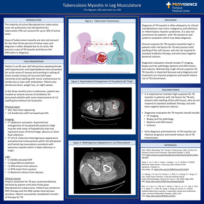
Tuberculous Myositis in the Leg Musculature
Tho Nguyen, MD
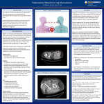
Introduction - The majority of active Mycobacterium tuberculosis cases are pulmonary, but extrapulmonary tuberculosis (TB) can account for up to 30% of active cases. Cases of tuberculosis myositis are rare and account for less than three percent of active cases and diagnosis is often delayed due to its rarity. We present a case of TB myositis and discuss the difficulties in diagnosis Case - Patient is an 82-year-old Vietnamese speaking female with hypertension and hyperlipidemia who presented for acute onset of nausea and vomiting in setting of three-month history of recurrent left lower extremity (LLE) swelling without systemic symptoms such as fever, weight loss, or night sweats. Patient reported several courses of antibiotics including sulfamethoxazole trimethoprim, clindamycin, and cephalexin in the past 3 months for presumed cellulitis with some improvement of LLE swelling but without full resolution. On physical exam, the patient had LLE tenderness with increased warmth. CT of LLE demonstrated extensive heterogeneous enhancement within the left gluteal and hamstring musculature with multiple irregular fluid collections present. The patient underwent drain placement for small fluid collections in left lateral thigh and hip. Atypical infections were considered including TB myositis and granulomatous myopathy. Work up was significant for mildly elevated CRP and a positive Quantiferon Gold test. AFB smears from abscess and sputum were negative as were bacterial cultures from the abscess. Treatment was recommended but not started until drain fluids grew Mycobacterium tuberculosis after the patient was discharged. On review of patient’s risk factors, her husband had latent TB but biggest risk was likely coming from an endemic area (Vietnam). The patient finished off 6 months of therapy with good response to treatment. Discussion – Diagnosis of TB myositis is often delayed as its clinical manifestations may mimic malignancy and infectious or inflammatory myositis syndromes. It is also not uncommon for patients presenting with TB myositis to not have systemic symptoms which may delay diagnosis. Clinical suspicion for TB myositis should be high in patients with risk factors for TB who present with swelling of the soft tissues, who do not respond to standard antibiotic therapy, and who have negative bacterial cultures. Diagnostic evaluation should include CT imaging, biopsy sent for pathology, bacteria and AFB smears, and cultures. Maintaining a high clinical suspicion for TB myositis is important because early diagnosis and treatment can improve prognosis and overall reduce risk of TB transmission.
-
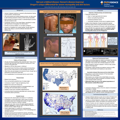
Not just a Biblical Disease: Hansen’s Disease (Leprosy) Oregon’s unique differential
Zach Taylor, Justin Jin, Andrea Roast, and Amy Dechet
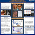
Background: Hansen’s disease (leprosy) is caused by a gram-positive, acid-fast bacilli called Mycobacterium leprae. It is very slow growing and has an indolent disease process. The disease is more prevalent in underdeveloped countries with poor access to care including Brazil, India, sub-Saharan Africa, and the Pacific Islands including Micronesia and the Philippines. However, it is becoming in creasingly common in the United States, specifically in areas with high levels of immigration from Micronesia. Case: Our patient was a 35-year-old Micronesian man who presented with 7 days of non-traumatic right hand pain with progressive swelling. He migrated to US from Micronesia in childhood and worked most of his life as a factory laborer. He often finds cuts and scrapes to his hands at home, though has no awareness of events that cause them. He does have a PMHx of prior hospitalization for burns/ scalds that he could not feel. His exam was pertinent for bilateral hand gangrene, prior loss of multiple digits, and multiple patches of neuropathy over his arms/ legs. He was diagnosed with osteomyelitis of the hand and started on appropriate IV antibiotics. However, the constellation of hand deformities, patchy areas of severe neuropathy, facial nerve findings and country of origin was concerning for leprosy. He was diagnosed over video consult with Hansen’s disease by a leprosy expert at the National Hansen’s Disease Center in Baton Rouge, LA. However the patient declined treatment and was lost to follow-up. Discussion: Hansen’s disease is largely a clinical diagnosis that has classic physical exam findings. There are nearly 250,000 new cases yearly in endemic regions, of which Micronesia is one. Interestingly, Micronesia is a part of the Compact of Free Association of the United States meaning that Micronesians can come/go to the U.S. at any time. Due to environmental and economic reasons, many Micronesians continue to migrate to the U.S. In fact, the Willamette Valley has one of the highest populations of Micronesians outside of Micronesia. There remains a lot of stigma surrounding this diagnosis related to prior perceptions that leprosy was highly communicable and untreatable. We now know this is not true, yet the stigma persists, and patients continue to suffer isolation, discrimination and hesitancy to see doctors. Conclusion: Hansen’s disease (leprosy) is rare, but it is here in Portland and the PNW, largely because of Micronesian migration patterns. Leprosy is a clinical diagnosis; endemic area + classic symptoms of hypopigmentation, patchy neuropathy and thickened nerves. There are Hansen’s Centers that can aid in diagnosis and guide treatment. Understanding stigma and correcting misinformation about futility of treatment and communicability may help overcome resistance to seeking care.
-
Don’t Poke the Bear: Pheochromocytoma Unmasked by COVID-10
Taylor Virtue and Andrea Roast
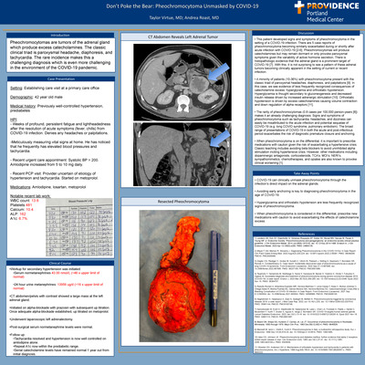
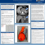
Introduction: Pheochromocytomas are tumors of the adrenal gland which produce excess catecholamines. The classic clinical triad is paroxysmal headache, diaphoresis, and tachycardia. The rare incidence makes this a challenging diagnosis which is even more challenging in the environment of the COVID-19 pandemic. Case Presentation: Setting: Establishing care visit at a primary care office Demographic: 42 year old male Medical history: Previously well-controlled hypertension, prediabetes HPI: • Weeks of profound, persistent fatigue and lightheadedness after the resolution of acute symptoms (fever, chills) from COVID-19 infection. Denies any headaches or palpitations. • Meticulously measuring vital signs at home. He has noticed that he frequently has elevated blood pressures and tachycardia. • Recent urgent care appointment: Systolic BP > 200. Amlodipine increased from 5 to 10 mg daily. • Recent PCP visit: Provider uncertain of etiology of hypertension and tachycardia. Started on metoprolol. Medications: Amlodipine, losartan, metoprolol Notable recent lab work: WBC count: 13.6 Platelets 481 Calcium: 10.4 ALP: 162 A1c: 6.7% Discussion: • This patient developed signs and symptoms of pheochromocytoma in the setting of a COVID-19 infection. There are 5 case reports of pheochromocy toma becoming similarly exacerbated during or shortly after acute infection with COVID-19 [2-6]. Pheochromocytomas will produce catecholamines but may remain dormant or only provoke paroxysmal symptoms given the variability of active hormone secretion. There is histopathologic evidence that the adrenal gland is a prominent target of COVID-19 [7]. With this, it is not surprising to see a pattern of these adrenal tumors becoming clinically apparent in the setting of current or recent infection. • A minority of patients (10-36%) with pheochromocytoma present with the classic triad of paroxysmal headaches, diaphoresis, and palpitations [9]. In this case, we see evidence of less frequently recognized consequences of catecholamine excess: hyperglycemia and orthostatic hypotension. Hyperglycemia is thought secondary to gluconeogenesis and decreased insulin release driven by increased adrenergic stimulation [10]. Orthostatic hypotension is driven by excess catecholamines causing volume contraction and down regulation of alpha receptors [11]. • The rarity of pheochromocytomas (0.8 cases per 100,000 person-years [8]) makes it an already challenging diagnosis. Signs and symptoms of pheo chromocytoma such as tachycardia, headaches, and dizziness can easily be misattributed to the acute infection and potential sequelae of COVID-19 (e.g. long COVID syndrome, pulmonary embolism). The broad range of presentations of COVID-19 in both the acute and post-infectious period exacerbates the risk of diagnostic premature closure and anchoring. • When pheochromocytoma is on the differential, it is important to prescribe medications with caution given the risk of exacerbating a hypertensive crisis. Classic teaching includes avoiding beta blockers to avoid uninhibited alpha stimulation inciting hypertensive crisis. However, other medications including dopaminergic antagonists, corticosteroids, TCA’s, MOI’s, NERI’s, sympathomimetics, chemotherapies, and opiates are also known to provike clinic worsening. Take Away Points: • COVID-19 can clinically unmask pheochromocytoma through the infection’s direct impact on the adrenal glands. • Avoiding early anchoring is key to diagnosing pheochromocytoma in the age of COVID-19. • Hyperglycemia and orthostatic hypotension are less frequently recognized signs of pheochromocytoma. • When pheochromocytoma is considered in the differential, prescribe new medications with caution to avoid exacerbating the effects of catecholamine excess.
-
A clot to Worry About: Venous Thromboembolism in Severe Ulcerative Colitis
Jin Yun, DO and Donald Lum
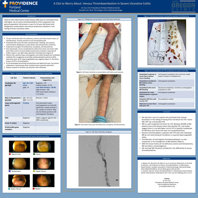
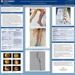
Introduction: Patients with inflammatory bowel disease (IBD) have an estimated three-fold higher risk of venous thromboembolism (VTE) compared to the general population. We present a case of a 19-year-old female with phlegmasia curelea dolens and massive pulmonary embolism in the setting of severe ulcerative colitis. Case Presentation : • 19-year-old female with iron deficiency anemia and three-month history of hematochezia, recently started on oral contraceptive pills. • Presented with acute left lower extremity pain, swelling, and cyanosis concerning for phlegmasia curulea dolens and compartment syndrome. • Underwent emergent thrombectomy, venoplasty, and fasciotomies. • Post-operatively, course complicated by obstructive shock and severe right heart failure due to massive bilateral pulmonary embolism requiring VA ECMO, heparin, and right sided aspiration thrombectomy (Figure 5). • Patient had a positive lupus anticoagulant and mildly elevated anti cardiolipin antibody (44.1) suggestive of possible antiphospholipid syndrome (APS), otherwise work up for hypercoagulability was negative (Figure 1). No family history of VTE or thrombophilia. • As she developed worsening hematochezia and abdominal pain, the patient underwent a colonoscopy which showed severe ulcerative pancolitis. • Patient received IV steroids and induction with Infliximab. Conclusions: • We describe a case of a patient who presented with massive thrombosis in the setting of having three risk factors for VTE: severe IBD, OCP use, and possible APS. • IBD is a well-recognized risk factor for VTE. Between 60-80% of IBD patients have active disease when they develop VTE, and studies suggest there is a six-fold higher risk for VTE in patients hospitalized for IBD flares than those that have non-hospitalized flares. • General recommendation is patients with VTE who have coexisting IBD do not need testing for hereditary or acquired hypercoagulable states. • Consideration of anticoagulant thromboprophylaxis is a vital component in the management of IBD patients (Table 2). • With her known history of iron deficiency anemia and hematochezia, IBD was likely a missed diagnosis. • We conclude IBD should be considered in the differential of venous thromboembolism.
-
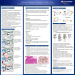
Immune Checkpoint Inhibitors: Friend or Foe
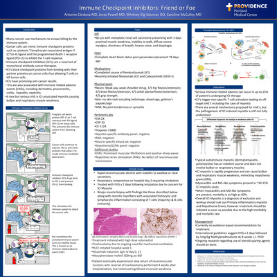
Antonio Cordova, MD; Jesse Powell, MD; Whitney Elg-Salsman, MD; and Caroline McCulley, MD
-
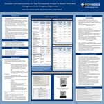
Evaluation and Implementation of a New Phenobarbital Protocol for Alcohol Withdrawal Management in the Emergency Department
Sarah J. Kim, Sara Clark, and Joshua L. Floyd
Background
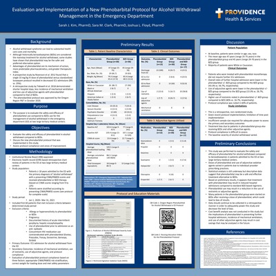
• Alcohol withdrawal syndrome can lead to substantial health care costs and mortality. • Although historically benzodiazepines (BZDs) are considered the mainstay treatment for alcohol withdrawal, some studies have shown that phenobarbital may be the safer and preferred alternative option. • Advantages of phenobarbital are its mechanism of action, more predictable pharmacokinetics, and greater therapeutic index. • A prospective study by Rosenson et al. 2012 found that a single 10 mg/kg IV dose of phenobarbital versus standardized lorazepam protocol resulted in decreased ICU admissions (8% vs. 25%). • A retrospective study by Tidewell et al. 2018 demonstrated shorter hospital stays, less incidence of mechanical ventilation and less use of adjunctive agents with phenobarbital compared to that of BZDs. • The phenobarbital protocol was approved by the Oregon Region P&T in October 2020.
Purpose
• The purpose is to evaluate the safety and efficacy of phenobarbital use compared to BZDs use for the management of alcohol withdrawal in the emergency department (ED) at two large tertiary medical centers.
Objectives
• Evaluate the safety and efficacy of phenobarbital in alcohol withdrawal compared to BZDs. • Discuss the new phenobarbital protocol that was implemented in this study. • Assess protocol compliance and areas of improvement.
Methodology
• Institutional Review Board (IRB)-approved • Electronic health record (EHR)-based retrospective chart review of patients in the ED at two large tertiary medical centers • Study population: • Patients ≥ 18 years admitted to the ED with the primary diagnosis of ‘alcohol withdrawal’ with a presenting CIWA/PAWSS scores and received phenobarbital or BZD therapy (based on CIWA scores ranging from 9 to >25) • Patients were stratified according to presenting CIWA/PAWSS scores (see protocol for complete scale) • Study period: • Jan 1, 2020– Mar 31, 2021 • Included first 60 patients that met inclusion criteria between determined study period • Exclusion criteria: • Allergy or hypersensitivity to phenobarbital or BZDs • Pregnancy • Documented history of acute intermittent porphyria, hepatic encephalopathy • Use of phenobarbital prior to admission as an anti- epileptic • Concomitant HIV medication use contraindicated with phenobarbital (Evotaz, Prezcobix, Tivicay, Doravirine, Genvoya, Stribild • Primary Outcome: ICU admission for alcohol withdrawal from the ED • Secondary Outcomes: Incidence of mechanical ventilation, use of restraints, use of adjunctive agents, and protocol compliance • Evaluation of phenobarbital protocol compliance based on three factors: appropriate CIWA/PAWSS risk stratification, correct weight for loading dose and met safety parameters
Discussion
Patient Population • At baseline, patients were similar in age, sex, race. • The mean age was 47 years (range 25-75 years) in the phenobarbital group and 45 years (range 29-70 years) in the BZD group. • Majority of patients were White or Caucasian. Clinical Outcomes • Patients who were treated with phenobarbital monotherapy did not require further ICU admission. • Overall rates of further hospital admission were lower in the phenobarbital +/- BZD group compared to the BZD group (43.3% vs. 56.7%, respectively). • Use of adjunctive agents were lower in the phenobarbital +/- BZD group compared to the BZD group (13.3% vs. 26.7%, respectively). • Higher use of restraints noted in phenobarbital +/- BZD group compared to BZD (40% vs. 16.7%, respectively). • Protocol compliance was noted in 60% of patients. Study Limitations • This is a retrospective, non-randomized study. • Given recent protocol implementation, limitation of time post implementation • Insufficient sample size required for adequate power to assess the primary and secondary outcomes • Treatment bias due to patients in phenobarbital group also receiving BZDs and other adjunctive agents. • Protocol compliance is difficult to assess. • The baseline severity of alcohol withdrawal varied.
Preliminary Conclusions
• This study was performed to evaluate the safety and efficacy of phenobarbital for alcohol withdrawal compared to benzodiazepines in patients admitted to the ED at two large tertiary medical centers. • Doses of phenobarbital and use of adjunctive sedative agents varied in patients due to individual provider prescribing practices. • Statistical analysis is still underway but descriptive data suggest that phenobarbital may be a safe and effective treatment alternative to BZDs. • Based on preliminary results, it appears that treatment with phenobarbital may result in reduced hospital admissions compared to standard BZD based regimens. • Phenobarbital use may result in a reduction in the use of restraints or adjunctive agents. • Many patients in the phenobarbital group were started on BZDs after receiving a dose of phenobarbital, which could lead to bias of results. • Data should continue to be collected in a retrospective manner in order to adequately power this study and decrease the level of bias. • Cost benefit analysis was not conducted in this study but the implications of phenobarbital in preventing further hospital admission, incidence of mechanical ventilation, and use of other adjunctive agents may result in cost savings that may be analyzed in the future.
Printing is not supported at the primary Gallery Thumbnail page. Please first navigate to a specific Image before printing.